

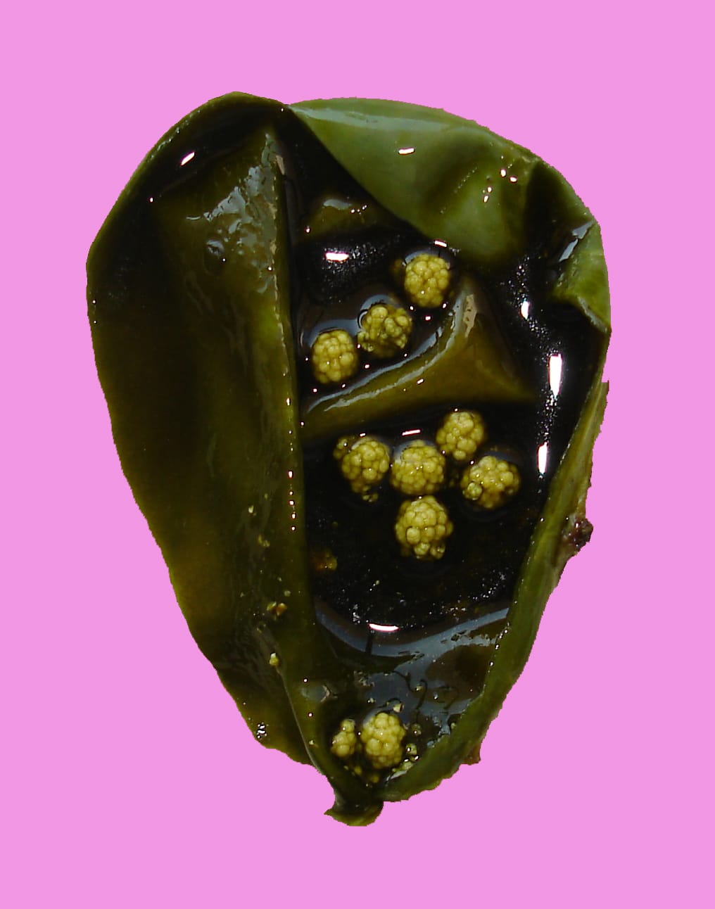
Gallbladder stones

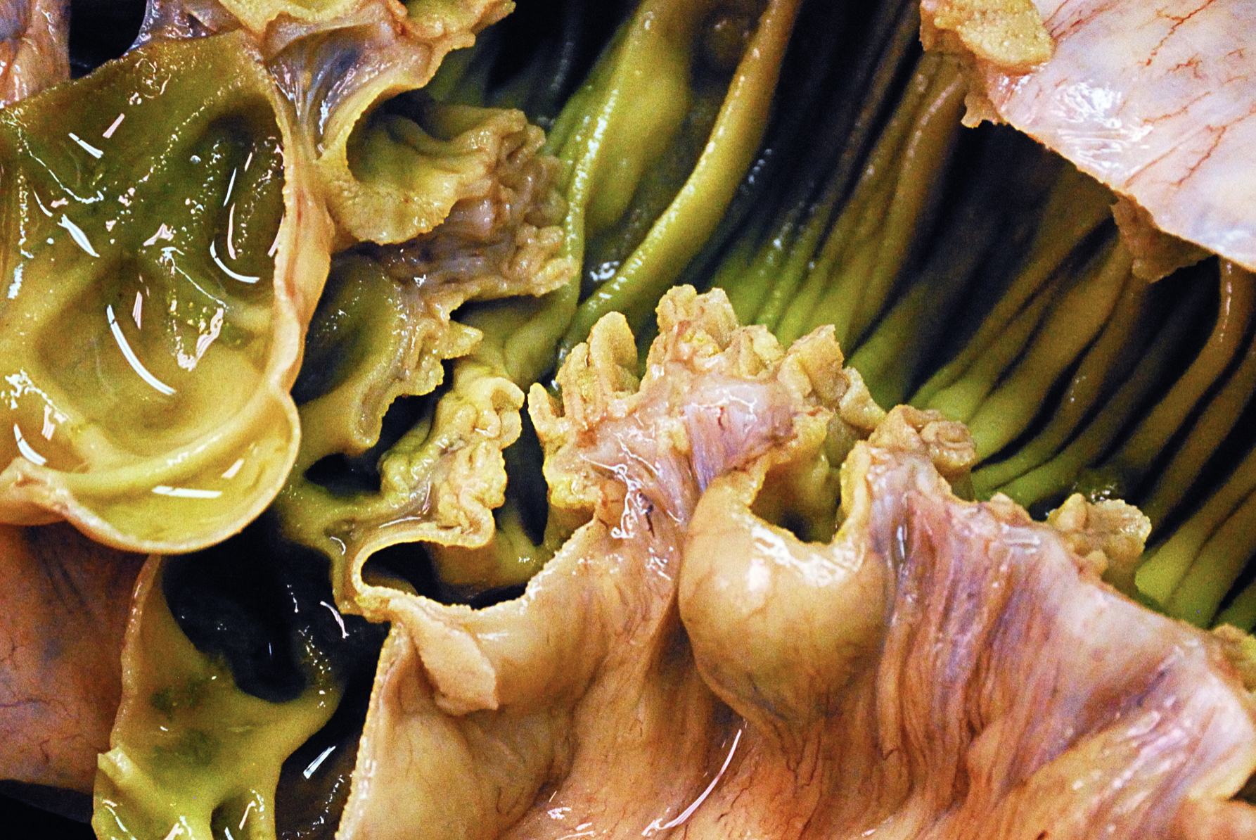
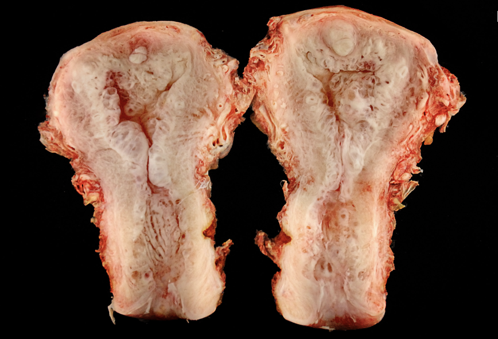
The large polyp on the left (central) is a cholesterol polyp, along with many smaller polyps on the surface. The large polyp on the right has a more pink than yellow color and represents a tubular adenoma, pyloric gland type. Cholesterolosis is seen throughout the mucosa.
Small intestine with multiple diverticuli present at the border of the mesenteric attachment. These show mucosal protusions through the muscular layers which are surrounded by muscularis mucosa and fat.
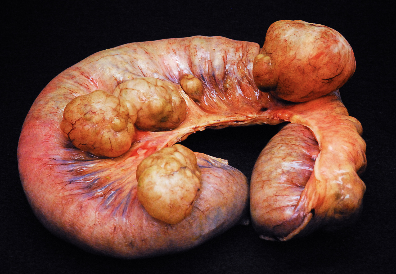
Small intestine with multiple diverticuli present at the border of the mesenteric attachment. These show mucosal protusions through the muscular layers which are surrounded by muscularis mucosa and fat.
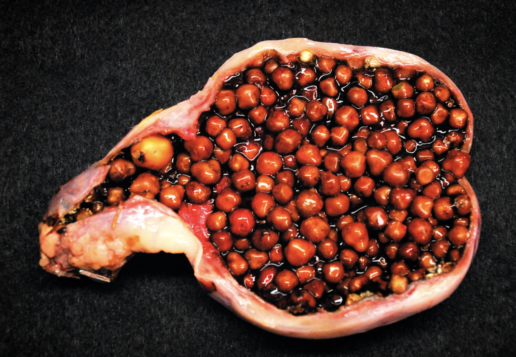
A gallbladder with marked Cholelithiasis.
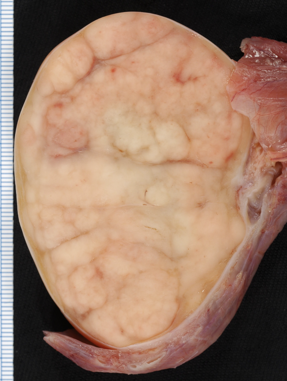
Endometrium with cystic atrophy.
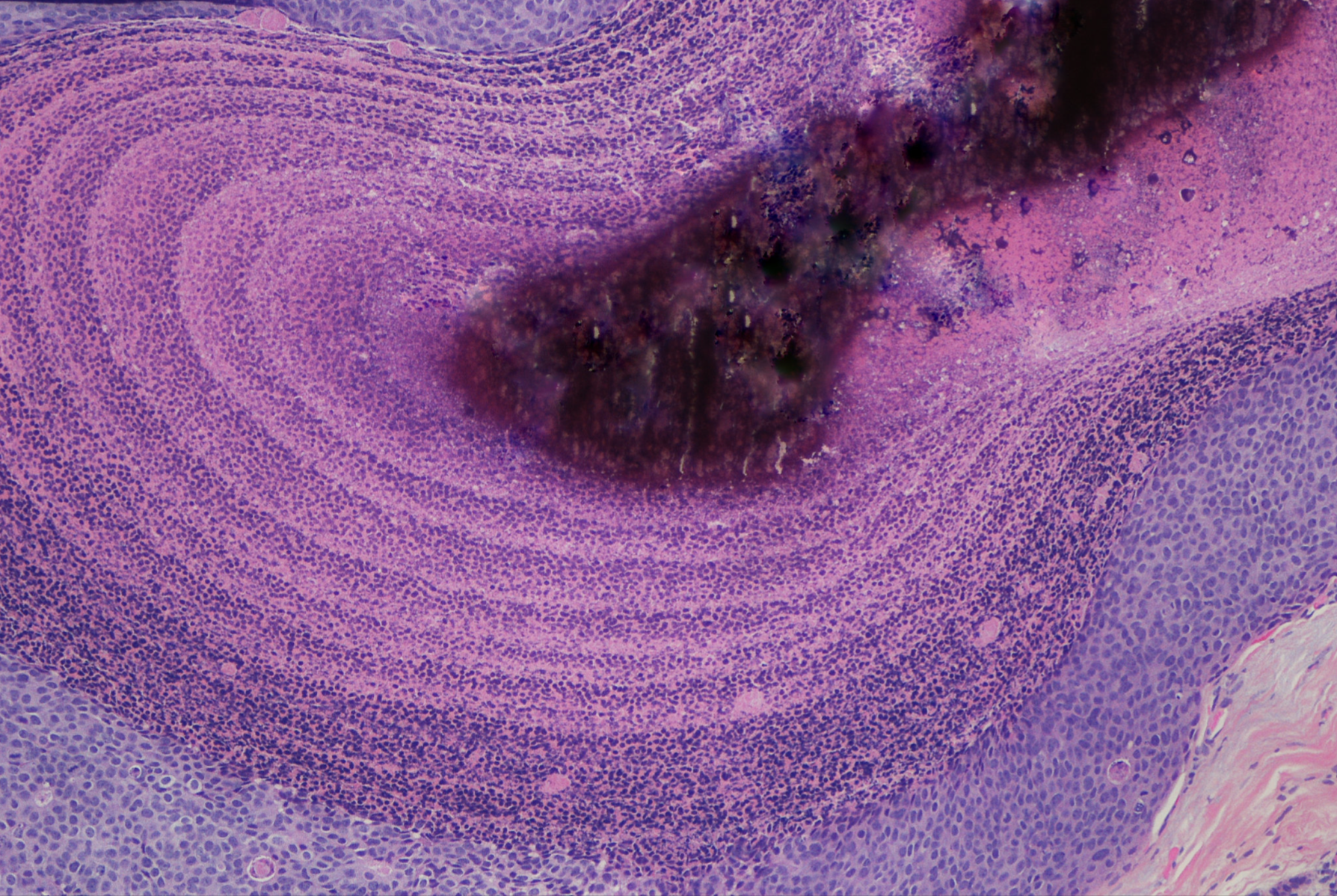
DCIS with calcification.
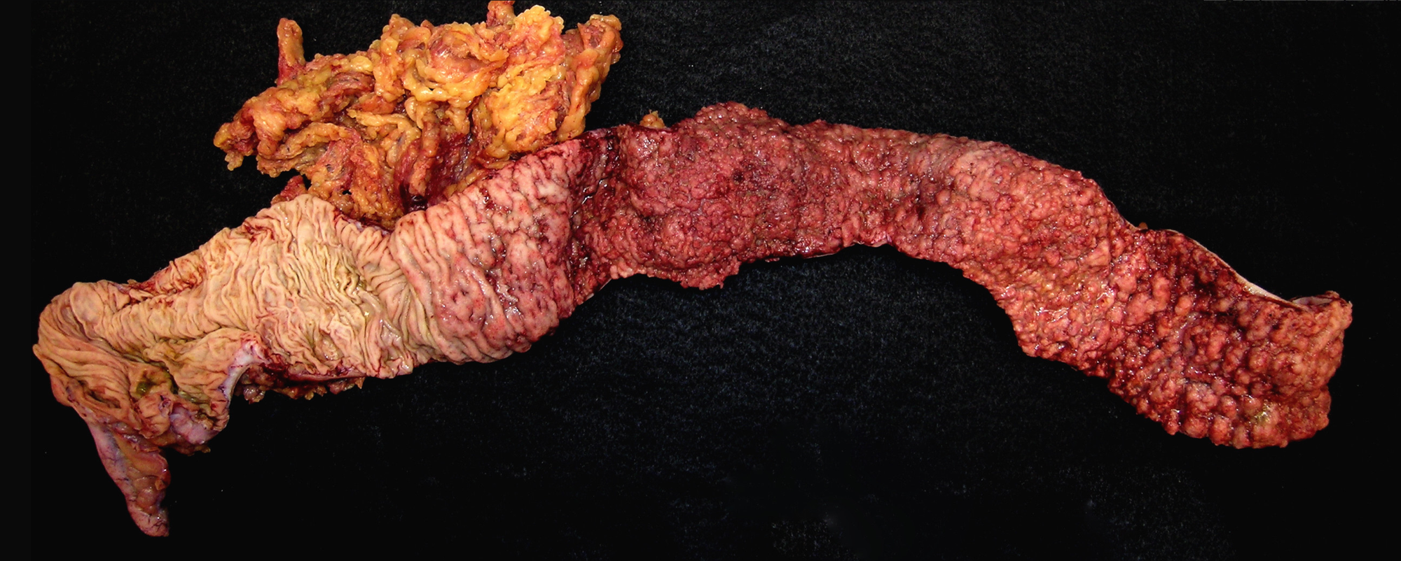
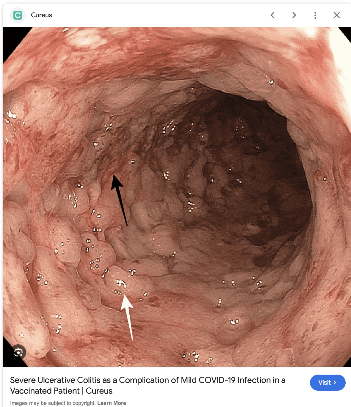
Acute severe (fulminant) ulcerative colitis, colectomy. Large ulcers with some fissuring are flanked by pseudopolyps. The fissures are limited to the submucosa and do not extend into the muscularis propria.
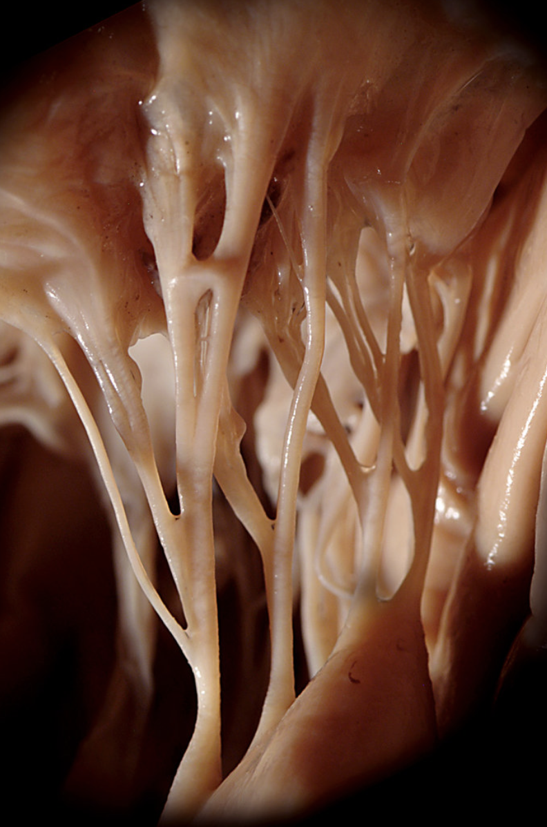
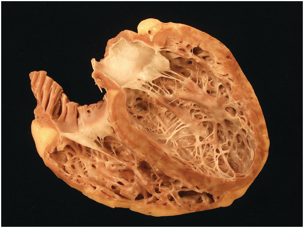
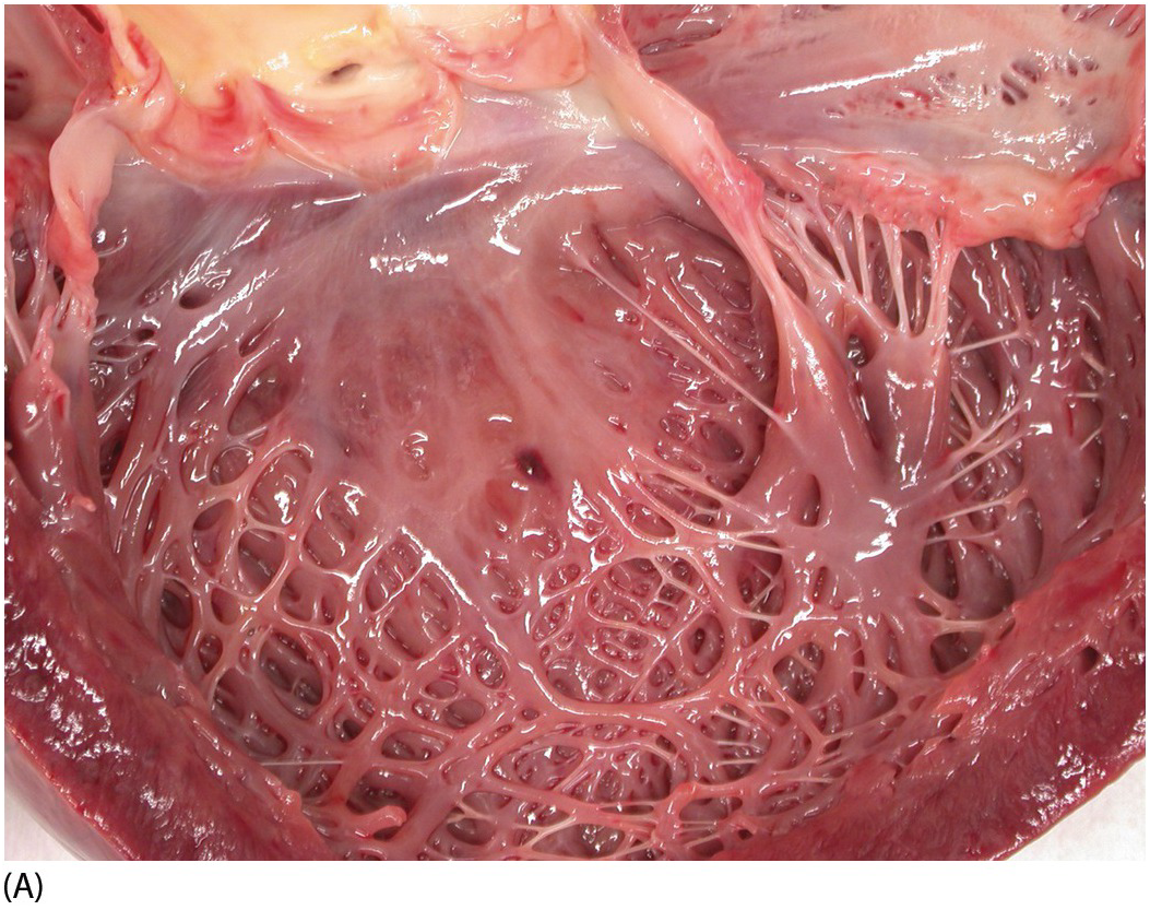
Myocarditis
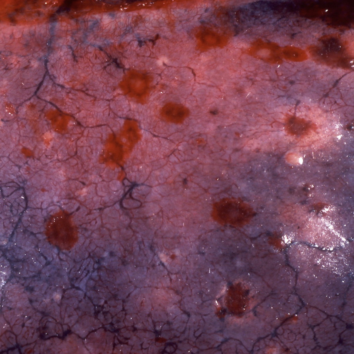
Liver damage
Liver damage
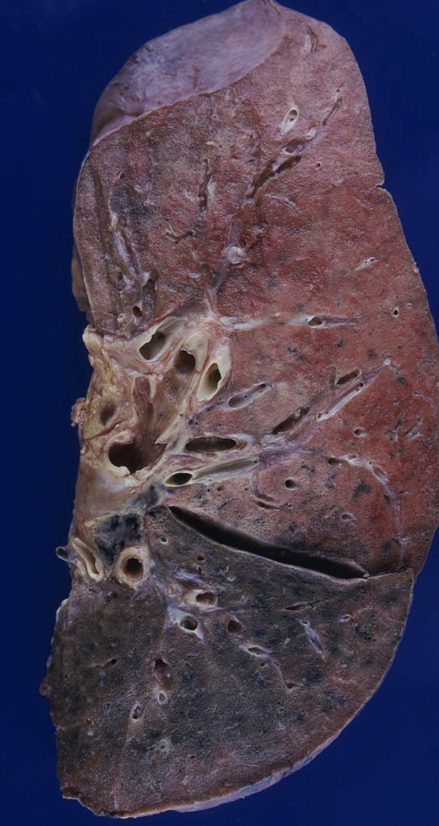
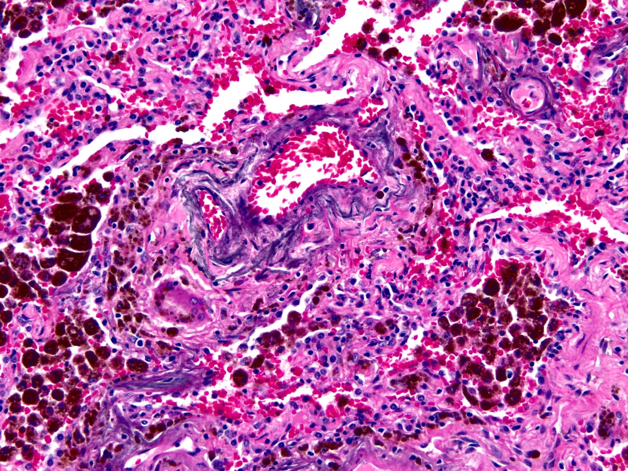
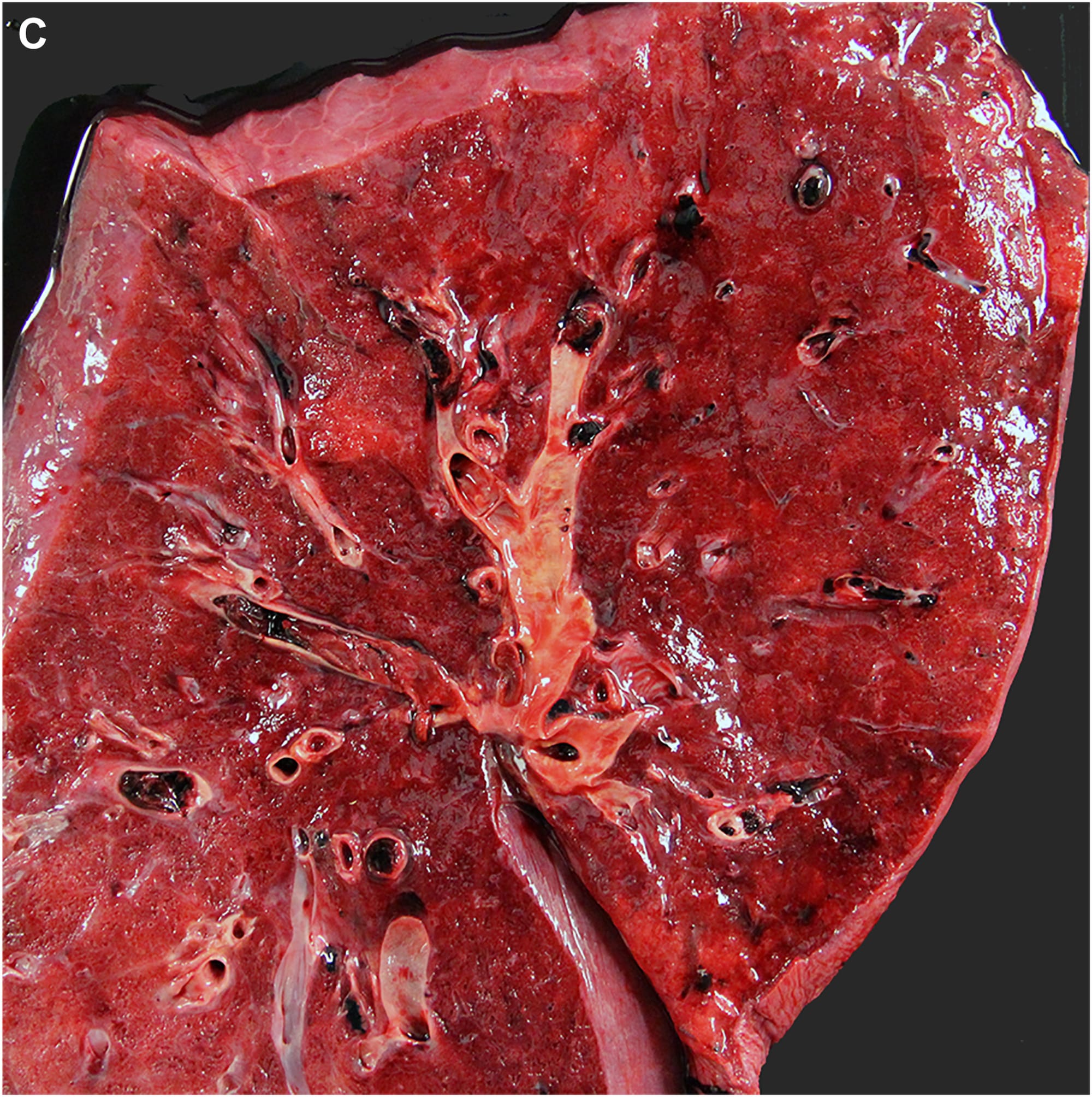
Lung Damage
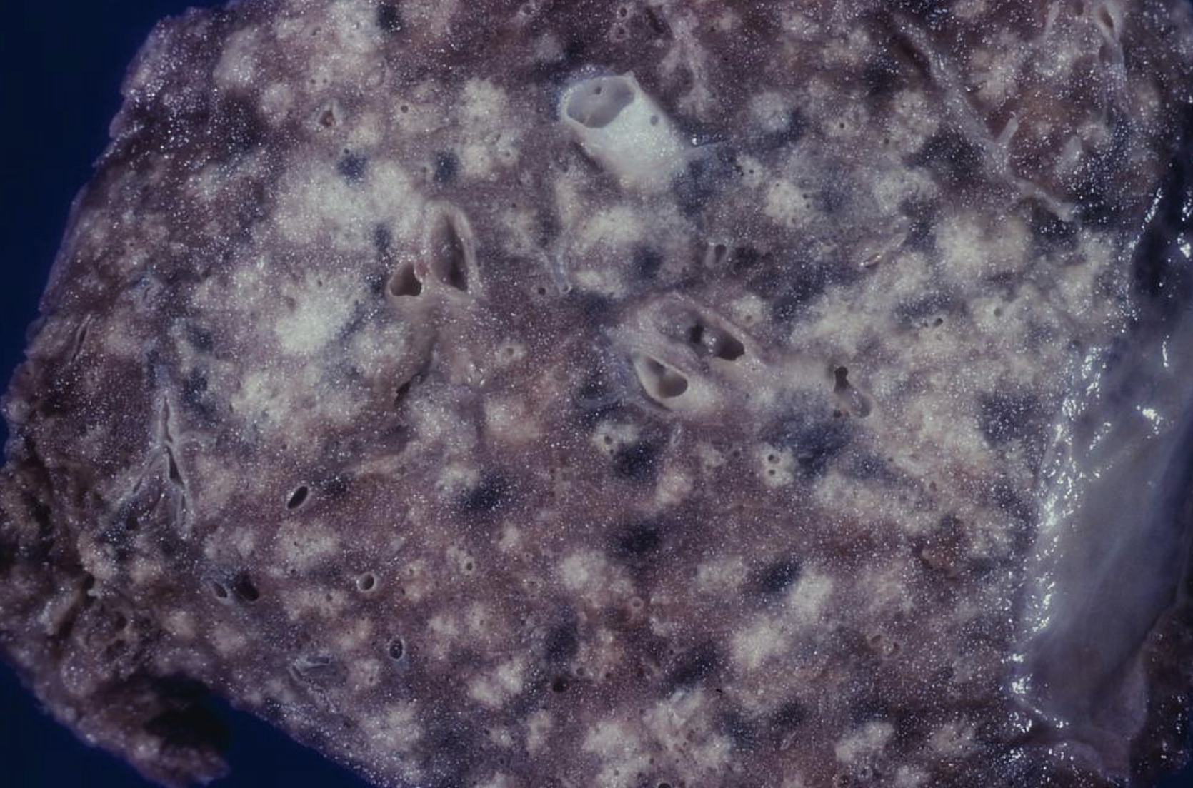
Lung Damage
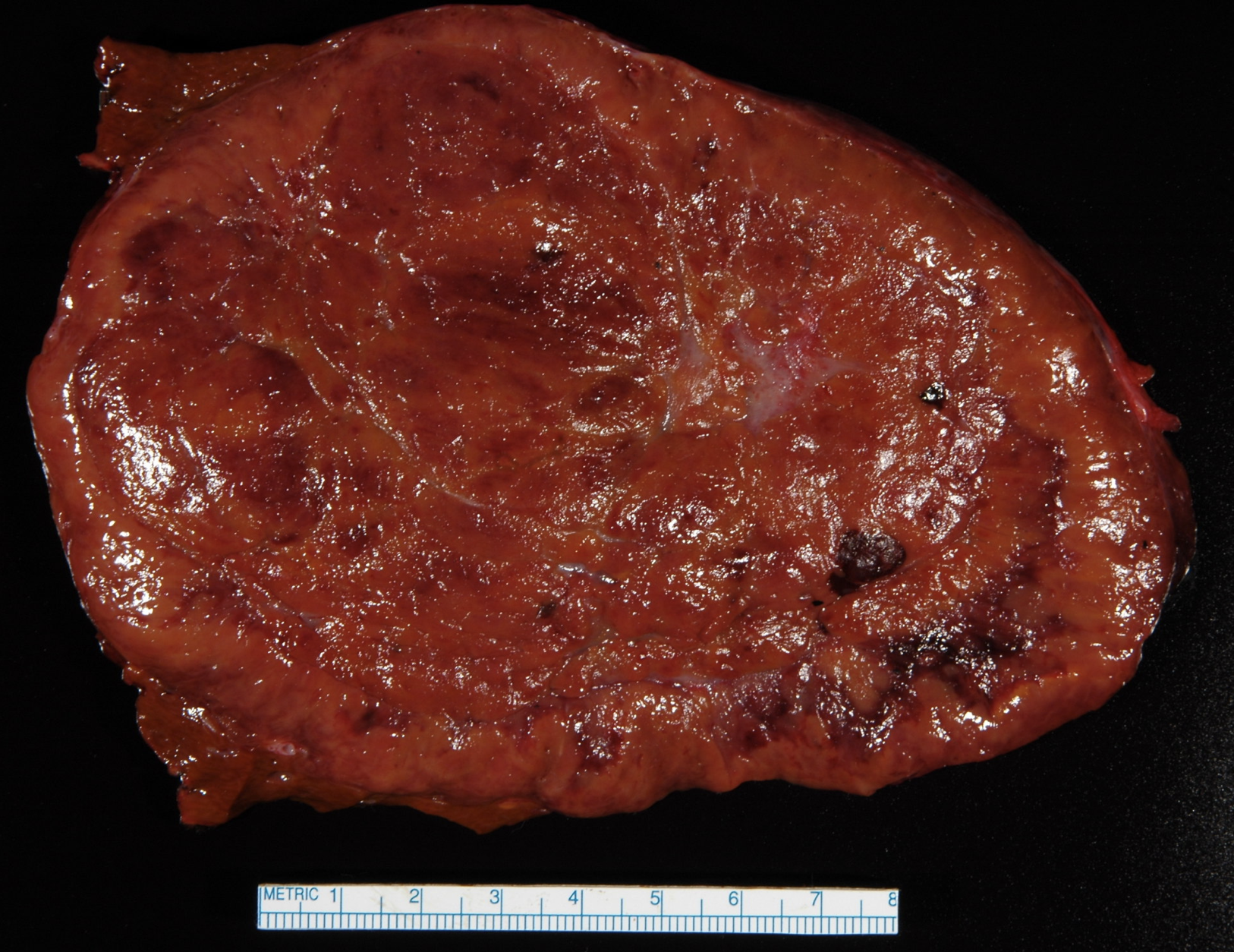
Varicocele
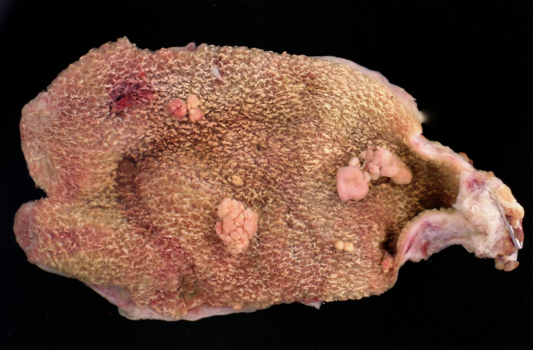
Polyps
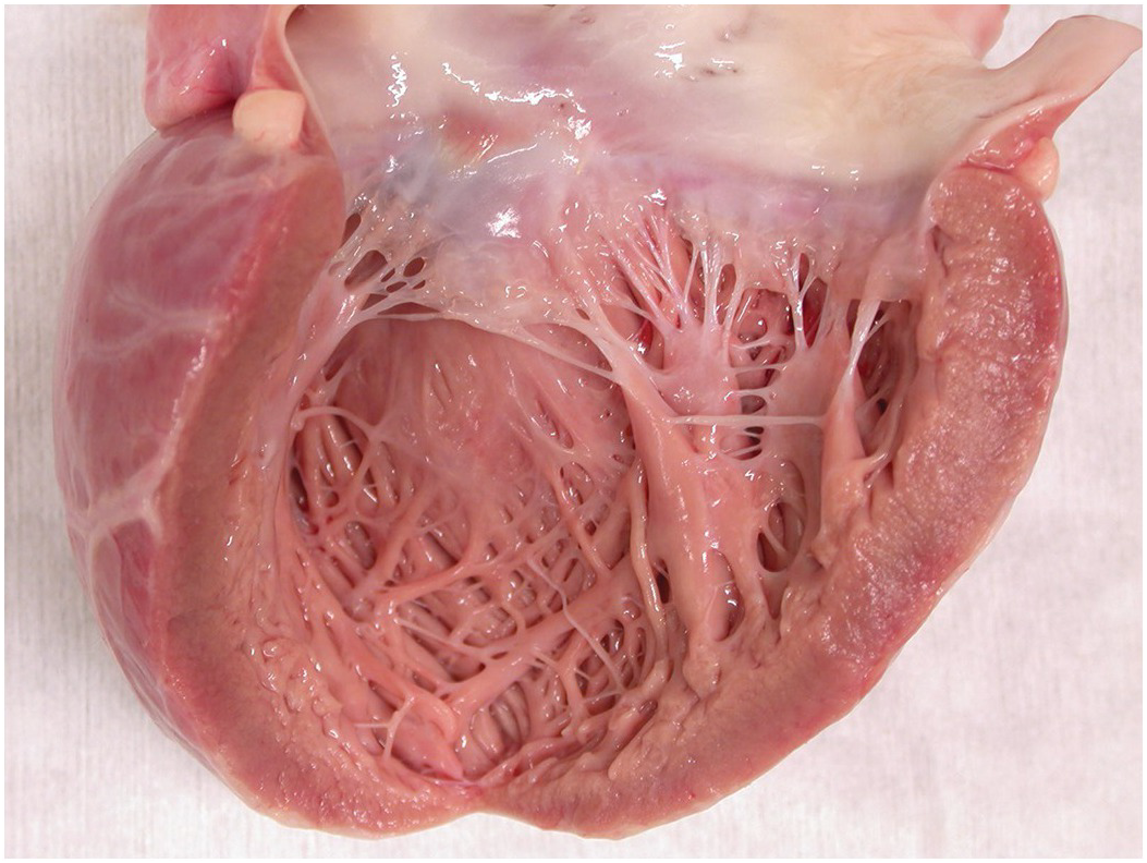
Myocarditis
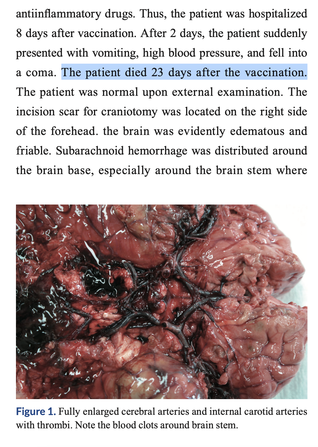


Discussion